Cone-beam CT scanning of
humans. Requires
careful geometry alignment and iso-centric C-Arms.
Radiotherapy
Positioning
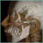
Dental
CT
Geometric accuracy in 3D for patient positioning and precise density resolution
for tumor localization.
The
use of tomographic 3D scanners for dental imaging is a newly
emerging modality.
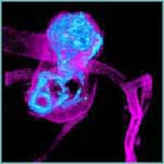
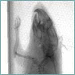
Scientific
CT
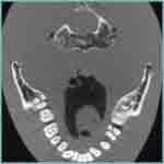
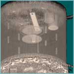
Industrial
CT
Cone
Beam CT scanning of small animals is
rapidly becoming the method of choice to study models of a
broad variety of human diseases.
Industrial scanning
differs in several aspects:
- samples exhibit strong eccentricity and
extreme dynamic range of the attenuation data
- spatial resolution is of high importance.
Exxim
has enhanced COBRA with a
practical iterative
algorithm, which is more
tolerant against input data imperfections than the
standard Feldkamp method. Such data imperfections
can have any nature, e.g. absorption non-linearities
or scanner geometry misalignment (COBRA V4.0 -
SAMARA).
In
under a minute, using only a PC, 2D projections can
be converted into a 512x512x512 3D image. This
software replaces current hardware- accelerated versions.
COBRA
software uses the full computational power of a
modern PC. It runs either on a single PC, or it can
be configured to run on a cluster of PC's (v.2
and higher); it
can also use modern graphics accelerators to do the
reconstruction (v.5
and higher).
COBRAsoftware
is implemented either as a stand-alone application
or as an SDK. In SDK mode, COBRA can start reconstruction
immediately after
getting the first projection from the CT scanner.
This allows the reconstruction and data acquisition
processes to run in parallel.